
Figure 5.
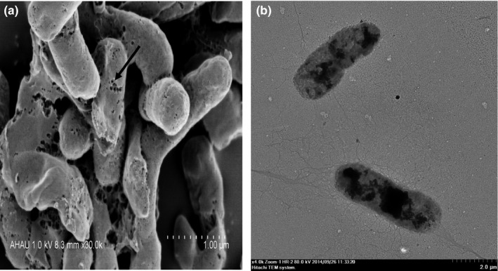
Vibrio alginolyticus strain 16‐3 BGs under electron microscopy. (a) Scanning electron micrograph (30,000 × ). Arrow indicates transmembrane tunnels. (b) Transmission electron micrograph (4,000 × ). The lysed cells show uneven and low electron density and retain the basic cell morphology