
Figure 4.
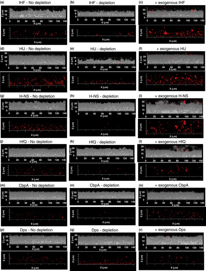
Depletion of NAPs from NTHI biofilm. Biofilm growth was initiated then maintained in the presence of one of the following: (a) respective preimmune serum (No depletion panels – a, d, g, j, m, p) (b) antiserum against the indicated NAP (depletion panels – b, e, h, k, n, q), (c) indicated NAP at 100 nmol/L (panels c, f, i, l, o, r) for 40 hr. Unfixed 40 hr biofilms were washed in PBS and incubated with anti‐IHF antiserum (a, b, c), anti‐HU antiserum (d, e, f), anti H‐NS antiserum (g, h, i), anti‐HfQ antiserum (j, k, l), and anti‐CbpA antiserum (m, n, o), and anti‐Dps antiserum (p, q, r) and then treated with goat anti‐rabbit IgG conjugated to AlexaFluor® 594 (NAPs – red). NTHI biofilms were visualized with DAPI (NTHI – gray). The biofilms were then imaged using a 63X objective on a Zeiss 510 Meta‐laser scanning confocal microscope (Carl Zeiss, Thornwood, NY). Note marked difference in biofilm height only upon depletion of IHF and HU and not upon depletion of non‐DNABII NAPs