
Figure 5.
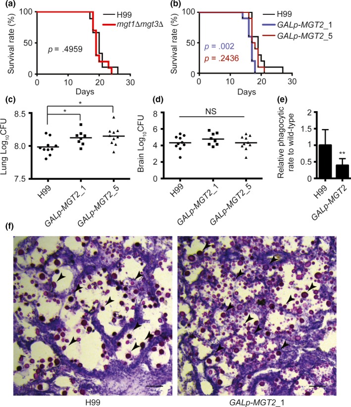
Analysis of C. neoformans Mg transporter mutants in mice. (a) Survival analysis of the wild‐type and mgt1Δ mgt3Δ strains. Twenty 4–6‐week‐old female C57BL/J mice were used for the analysis. Overnight fungal cell cultures were washed twice with PBS and diluted with PBS. A PBS suspension containing 105 cells of the wild‐type or mgt1Δ mgt3Δ strain were used for the infection of mice. Animal survival rates were recorded. (b) Survival analysis of the wild‐type and GAL7p‐MGT2 strains. Twenty 4–6‐week‐old female C57BL/J mice were used for the analysis. Overnight YPGal fungal cell cultures were washed twice with PBS and diluted in YPD medium. The YPD cultures were incubated for 6 hr to repress the GAL promoter. Cells were then washed and diluted. A PBS suspension containing 105 cells of the wild‐type or mgt1Δ mgt3Δ strain were used for infection in mice. Animal survival rates were recorded. (c) Fungal burden analysis in the lungs. Two independent GAL7p‐MGT2 strains were used to infect mice as described in Figure 5b. Fourteen days post infection, mice were killed, and CFUs from the lung tissues were analyzed. (d) Fungal burden analysis in the brain. The brain fungal burden tests were performed as described in Figure 5c. ANOVA was used for statistical analysis (*p < .05). (e) Phagocytic assay of mgt2 mutant. YPGal fungal cell cultures were washed twice with PBS, and then mixed with RAW264.7 cells in DMEM medium supplemented with 10% FCS. Extracellular fungal cells were washed with PBS buffer. Internalized and membrane‐bound fungal cells were counted. Relative phagocytic rates were calculated. The graph represents three biological replicates, and over 150 macrophages were counted for each replicate. The Student's t‐test was performed to determine statistical significance (**p < .01). (f) Histopathological analysis of C. neoformans‐infected lung tissues. Lung tissues from H99‐ and Gal7p‐MGT2‐infected mice were isolated, fixed, and stained with mucicarmine or H&E. Arrows indicates strained C. neoformans cells