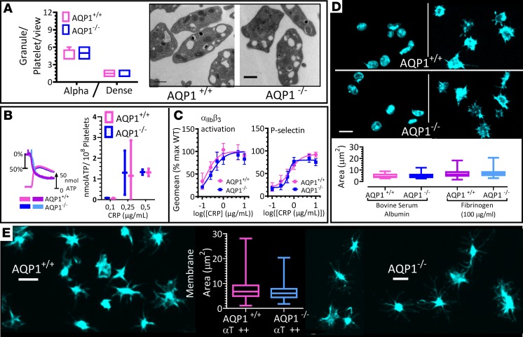
Figure 2. Ablation of AQP1 does not alter integrin activation or secretion in mouse platelets.
(A) Transmission electron microscopy of AQP1+/+ and AQP1–/– mouse platelets. Representative images are shown, and granule count was determined. (B) Ablation of AQP1 had no effect on platelet-dense granule release. AQP1+/+ and AQP1–/– mouse platelets were stimulated with 0.5 μg/ml collagen-related peptide (CRP), and ATP secretion was assessed by luminometry. Representative trace and graph with levels of secretion (mean ± SEM). Blue and cyan tracings of chart show percentage aggregation and the simultaneous ATP release recorded in AQP1+/+ mouse platelets, respectively. Corresponding tracings for AQP1–/– platelets are shown in light and deep magenta. (C) Washed mouse platelets (5 × 10⁷/ml) from AQP1+/+ or AQP1–/– mice were stimulated for 10 minutes with a range of concentrations of CRP in the presence of 1 mM CaCl2. Integrin αIIbβ3 activation and P selectin exposure were measured by flow cytometry. The geometric mean of the fluorescence intensity was determined, and data are shown as the percentage of maximal control (AQP+/+) response. Curves were fitted by F test. (D) Washed platelets from wild-type (AQP1+/+) or AQP1-null (AQP1–/–) mice allowed to adhere to BSA-coated (2%) or fibrinogen-coated (100 μg/ml) surfaces and stained for actin (FITC-phalloidin). Quantification of spreading (surface area) in box-and-whisker plots. (E) Platelets adherent to BSA were stimulated with 1 U/ml thrombin and spreading analyzed as in D. Statistical significance was determined by 2-way ANOVA and Bonferroni post hoc test (C) and by Wilcoxon signed-rank test (A, B, and E). Scale bar: 500 nm (A); 3 μm (D and E). Data were from 6 independent experiments.

