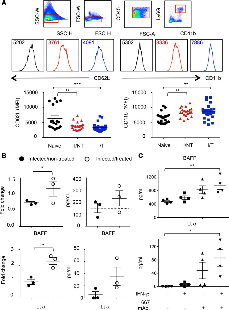
Figure 7. Activation of splenic and BM-isolated neutrophils.
(A) Expression of CD11b and CD62L. Spleen cells from naive, I/NT, and I/T mice were isolated at day 8 p.i. and were analyzed by flow cytometry for assaying cell surface expression of CD11b and CD62L. The data represent 5 independent experiments, with at least 18 mice per group. Data are expressed as mean ± SEM. (B) Expression and protein release of BAFF and LTα by neutrophils. Neutrophils from naive, I/NT, and I/T mice were sorted from spleens at day 8 p.i. and assessed for cytokine expression or protein release. Cytokine expression (left) was assessed by RT-qPCR normalized to β-actin. The data show fold changes in cytokine expression by neutrophils from I/NT and I/T mice as compared with naive mice and are representative of 3 independent experiments, with 8–10 mice per group. Protein release (right) was assessed by ELISA in supernatants of sorted neutrophils cultured at a density of 2 × 10⁵ cells/well for 24 hours. The data show BAFF and LTα release by neutrophils from I/NT and I/T and are representative of 3 independent experiments, with 8–10 mice per group. The dashed line represents the level of BAFF released by neutrophils sorted from naive mice. No LTα release was detected from neutrophils sorted from naive mice. (C) BAFF and LTα release by BM-isolated neutrophils. BAFF and LTα release was assessed by ELISA in supernatants of neutrophils isolated from BM of naive mice (>95% purity) and cultured for 24 hours in 667 mAb–coated 24-well plates at a density of 2 × 10⁶ cells in 500 μl medium. Experiments were done in the presence and in the absence of the proinflammatory cytokine IFN-γ (100 ng/ml). 667 mAb–noncoated plates were used as control. The data represent 4 independent experiments. Data are expressed as mean ± SEM. Statistical significance was established using a parametric 1-way ANOVA test with a Bonferroni correction (A and C) or a paired Student’s t test (B) (*P < 0.05; **P < 0.01; ***P < 0.001).

