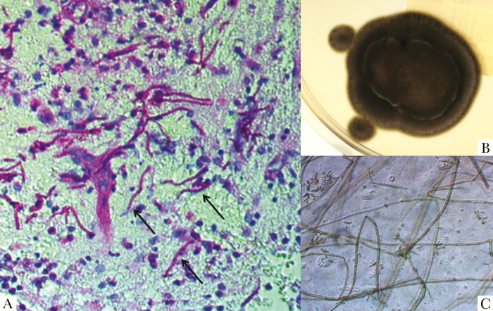
Figure 2.
A, Numerous purple-stained hyphae (black arrows) on Periodic acid Schiff (PAS) stain of brain tissue (magnification x40). B, Black velvet-like colonies of Fonsecaea monophora on Sabouraud Glucose Agar after 12 days of incubation at 25°C. C, Micromophology of colony fragments on lactophenol cotton blue stain (LCBS) with septate hyphae, conidiophores, and many solitary conidias (magnification x400).

