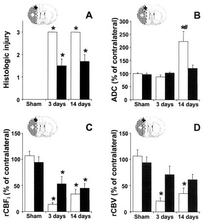
Figure 7.
Histologic tissue injury (A), relative ADC (B), rCBFi (C), and rCBV (D) in the ischemic core (white bars) and in ipsilesional M1/S1fl (black bars) at 3 and 14 days after stroke. *, P < 0.05 vs. sham-operated rats; #, P < 0.05 vs. 3 days after stroke. ROIs in the ipsilesional M1/S1fl (black cross) and ischemic core (white cross) are schematically represented on inserted outlines of a coronal rat brain section. The ischemic lesion is stippled.

