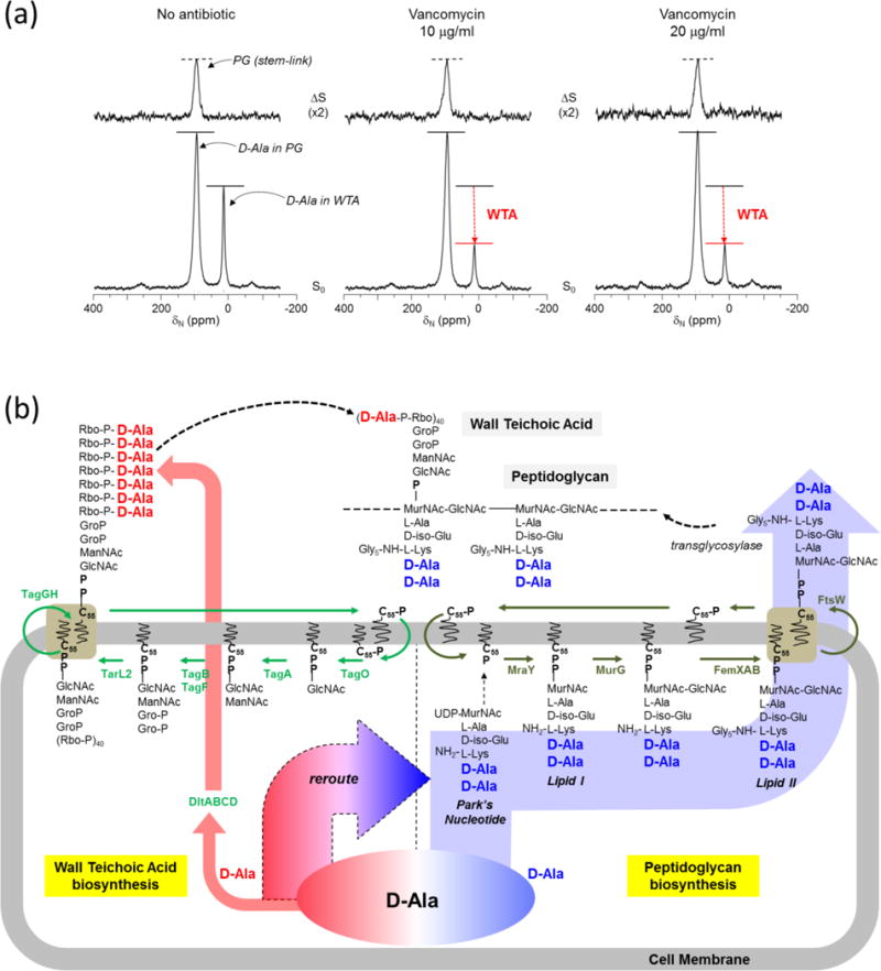
Figure 4. Vancomycin-treated S. aureus show inhibition of WTA biosynthesis preceding PG biosynthesis.

a, D-[15N]Ala and L-[1-13C]Lys labeling of S. aureus PG and WTA in presence of alanine racemase inhibitor alaphosphin (5 ug/ml). 93-ppm peak intensities in the ΔS spectra (top) of 15N{13C} REDOR at 1.6 ms show that alaphosphin improves D-[15N]Ala incorporation into PG. The addition of vancomycin did not affect the PG stem-link density; however, the S0 spectra show a large reduction in the D-[15N]Ala incorporation into WTA (16 ppm). b, Schematic representation of WTA and PG biosyntheses with bactoprenol-phosphate (C55-P) as the central lipid transporter. Glycopeptide antibiotics prevent regeneration of the lipid transporter and thereby inhibit both PG and WTA biosyntheses. However, as shown in Fig. 4a D-[15N]Ala incorporation into WTA (red arrow) was inhibited in vancomycin-treated S. aureus by rerouting all available D-[15N]Ala into maintaining the PG biosynthesis (blue arrow). Hence, the vancomycin inhibition of D-[15N]Ala incorporation into the WTA biosynthesis precedes interference of the PG biosynthesis.
