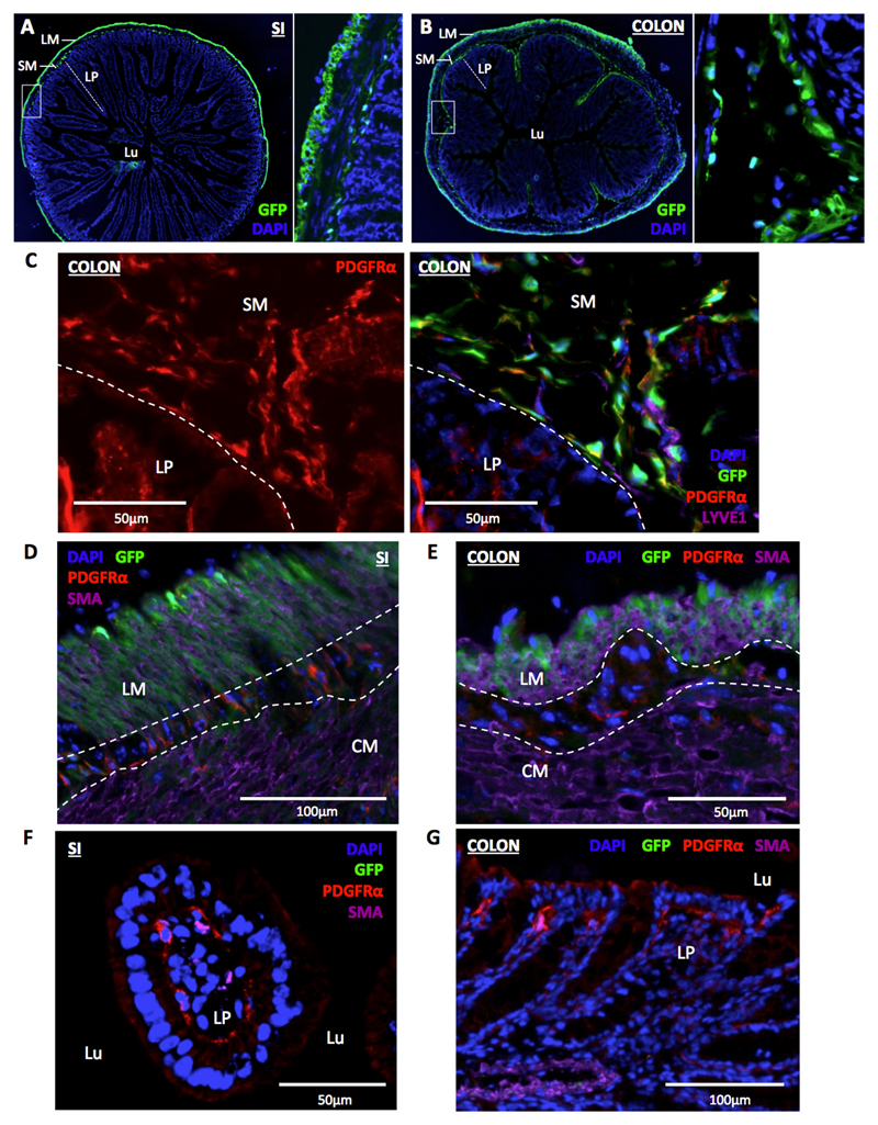
Figure 5. Ackr4+ fibroblasts populate the intestinal submucosa.
(A-B) Representative fluorescent microscopic images showing the location of GFP expression (green) in transverse sections of (A) small intestine (SI) and (B) colon of Ackr4gfp/+ (Ackr4tm1Ccbl) mice co-stained with DAPI (blue). The image on the right of each panel is a magnified version of the region shown in the box on the image to its left. LM, longitudinal muscle; SM, submucosa; LP, lamina propria; Lu, lumen. (C) Representative immunofluorescent microscopic image of section of colon from Ackr4gfp/+ mouse immunostained with Abs against PDGFRα (red) and LYVE1 (purple), and counterstained with DAPI (blue). Left, PDGFRα (red) only; right, composite image of all colours. The submucosa (SM) and lamina propria (LP) are indicated and separated by the dotted line. (D-G) Representative immunofluorescent microscopic images of sections of (D, F) SI or (E, G) colon of Ackr4gfp/+ mice immunostained with Abs against PDGFRα (red) and αSMA (purple), and counterstained with DAPI (blue). In D and E, longitudinal muscle (LM) and circular muscle (LM) layers are labeled and their boundaries marked with dotted lines; in F and G, lamina propria (LP) and lumen (Lu) are marked. Scale bars are shown on the images. Images are representative of at least two individual experiments each involving two or more mice.

