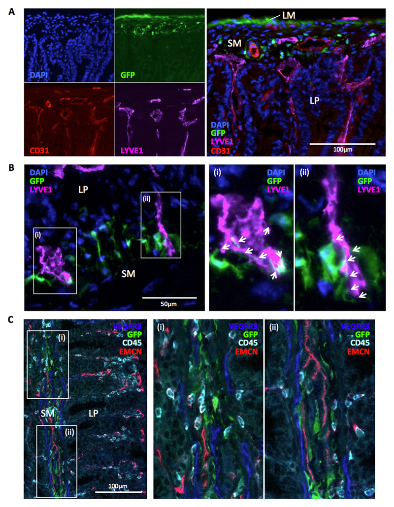
Figure 8. Intestinal Ackr4+ fibroblasts interact with the submucosal vasculature.
(A-B) Representative images captured by fluorescence microscopy from sections of small intestine from Ackr4gfp/+ (Ackr4tm1Ccbl) mice immunostained with fluorescent Abs against (A) CD31 (red) and LYVE-1 (purple) or (B) LYVE1 only (purple), and co-stained with DAPI (blue). In A, the images in the left panel show individual colours; a composite image is shown in the right panel. In B, images in (i) and (ii) show high power views of the areas highlighted in the left panel, with arrows indicating sites of physical interaction between LYVE1+ cells and GFP+ cells. LM, longitudinal muscle; SM, submucosa; LP, lamina propria. (C) Image of section of colon from Ackr4gfp/+ mouse immunostained with Abs against VEGFR3 (blue), CD45 (cyan) and blood vessel protein, endomucin (EMCN, red). Right panels show high power views of the areas (i) and (ii) highlighted in left panel. Scale bars are shown. Images are representative of those taken from at least three independent experiments.

