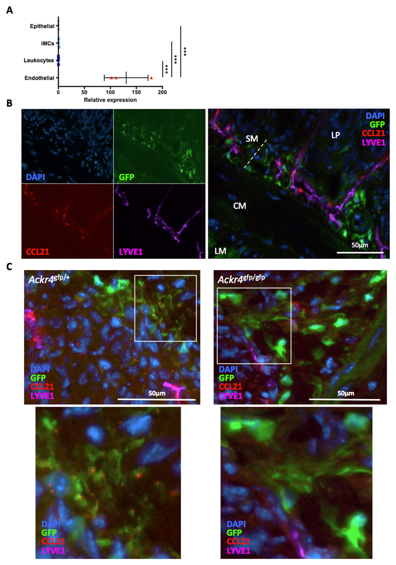
Figure 9. ACKR4-dependent association of LEC-derived CCL21 with submucosal Ackr4+ fibroblast.
(A) cDNA was prepared from RNA extracted from endothelial cells (CD45-CD31+), leukocytes (CD45+), intestinal mesenchymal cells (iMCs; CD45-GP38+CD31-) and epithelial cells (CD45-EPCAM+) that had been FACS-sorted from the small intestine. Expression of Ccl21 was analyzed by QPCR. Mean expression by iMCs is set to 1. Data points from individual mice are shown, along with means (±1SD). ***p< 0.001; one way ANOVA. (B-C) Representative images of sections of small intestine from (B, C) Ackr4gfp/+ or (C) Ackr4-deficient Ackr4gfp/gfp (Ackr4tm1Ccbl) mouse immunostained with Abs against CCL21 (red) and LYVE1 (purple), and co-stained with DAPI (blue). In B, the images in the left panel show individual colors; a composite image is shown in the right panel. LM, longitudinal muscle; CM, circular muscle; SM, submucosa; LP, lamina propria. In C, lower panels show high power views of the areas highlighted in the panel above. Scale bars are shown. Images are representative of two or more individual experiments.

