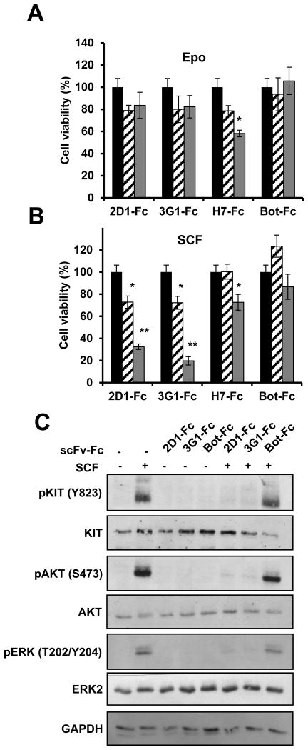
Figure 3. Effect of anti-KIT D5 domain scFv-Fc 2D1 and 3G1 on UT-7/Epo cell line.
A, B, UT-7/Epo cells were incubated 4 days in Epo (A) or CHO-KL supernatant (B) in the presence of anti-KIT antibodies or control antibody as indicated at 5 μg/mL (dashed) or 50 μg/mL (grey). Cell viability was measured by MTS assay and is expressed in % compared to non-treated cells (black). Data were analyzed with a Kruskal Wallis test. Differences were considered significant when p < 0.05: *, p < 0.05; **, p < 0.01, versus non treated cells. C, UT-7/Epo cells were starved overnight in serum-free medium and incubated with scFv-Fc (10 μg/mL) before 5 min stimulation with SCF (100 ng/mL). KIT, AKT and ERK1/2 phosphorylation were analyzed by Western blot with phospho-specific antibodies. Total KIT and ERK2 levels were visualized after stripping the membranes and reprobing with anti-KIT, anti-AKT and anti-ERK2 antibodies.

