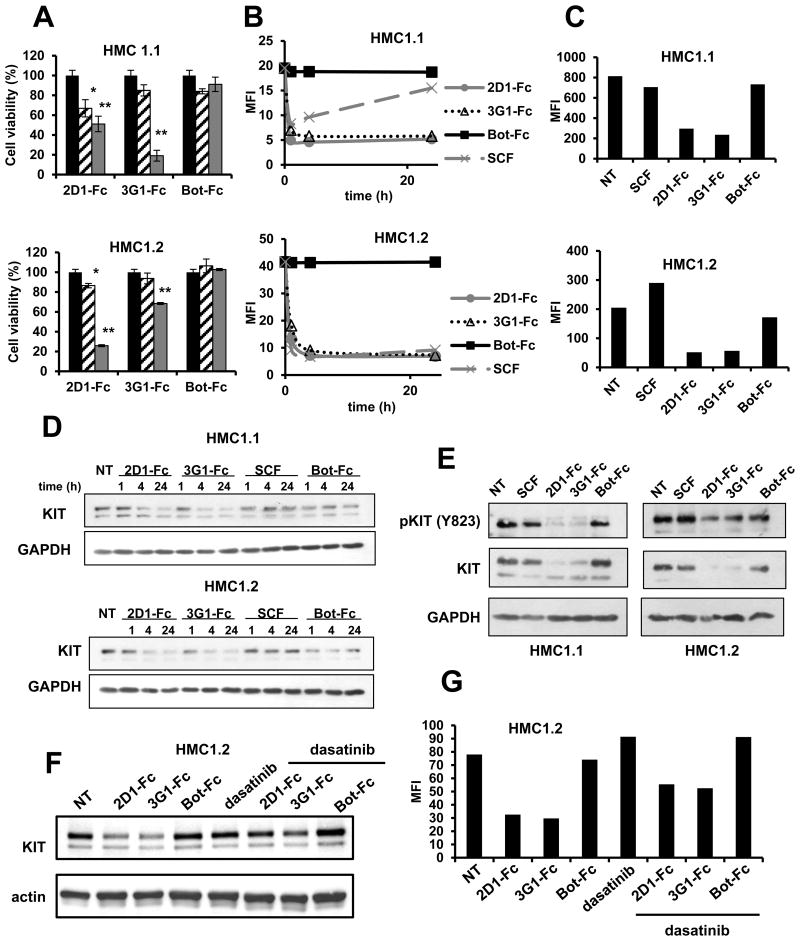
Figure 5. Oncogenic KIT modulation by anti-KIT D5 scFv 2D1 and 3G1 in HMC1 cell lines.
A, HMC1 cell lines were incubated for 7 days in IMDM 1% FCS in presence of anti-KIT (2D1 and 3G1-Fc) or control scFv-Fc antibody (Bot-Fc) at 5 μg/mL (dashed) or 50 μg/mL (grey). Cell viability is expressed in % compared to non-treated cells (black). Data were analyzed with a Kruskal Wallis test. Differences were considered significant when p < 0.05: *, p < 0.05; **, p < 0.01, versus non treated cells. (B-G) HMC1 cells were incubated in IMDM 10% FCS in presence of scFv-Fc antibodies (10 μg/mL), SCF and dasatinib (1 μM) as indicated for 1, 2 and 24 hrs (B, D), 72 hrs (C, E) or (F, G) 4 hrs. Treated cells were analyzed by FACS using 104D2 anti-KIT antibody (B, C, G) or by Western blot using anti-KIT and GAPDH antibodies (D, E, F). Representative data of 3 independent experiments each time.

