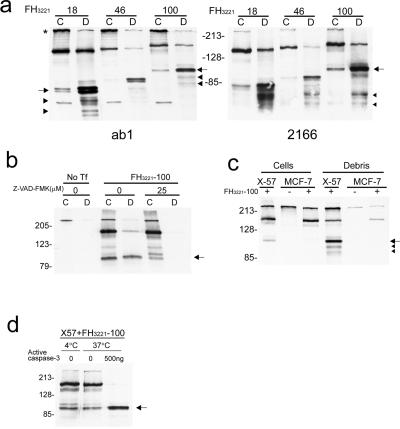
Figure 1.
Western blots show caspase 3 cleavage of htt in transfected cells and in mouse brain. (a) X57 cells were transfected with FLAG-htt cDNAs (FH3221) with 18, 46, or 100 glutamines. Blots are from different gels run in parallel and probed with anti-htt antisera ab1 (Left) and 2166 (Right).The N-httcasp3 fragment varies in size from about 80–100 kDa (arrows) depending on polyglutamine length in htt and is present in cells (C) and in debris (D). The N-httcasp3 fragment appears as a doublet (left arrow). Proteolytic fragments of the N-httcasp3 fragment are identified at the level of the arrowheads. The uncleaved protein expressed from FH3221 migrates between 140 and 160 kDa depending on polyglutamine length in htt. Native mouse full-length htt (*) and a small mouse N-htt fragment is seen in cells in blot on left. (b) X57 cells were transfected with FH3221-100 or not transfected (No Tf) and cells (C) and debris (D) were assayed for htt expression with Ab1. In the transfected cells, treatment with Z-VAD-FMK (25 μM) attenuates the level of the N-httcasp3 fragment (arrow), and there is an increase in the level of a precursor fragment. The N-httcasp3 fragment is not present in debris (D) from cells treated with Z-VAD-FMK. (c) X57 cells and MCF-7 cells were transfected with FLAG-htt cDNA FH3221-100. The N-httcasp3 fragment (arrow) appears in cells and debris of X57 cells but is not present in the caspase 3 deficient MCF-7 cells. Proteolytic products of the N-httcasp3 fragment are identified at arrowheads in the debris of X57 cells. The blot was probed with Ab1. Native mouse htt is at the top. (d) Cleavage of human htt by active caspase 3. Protein extracts from X57 cells transfected with mutant htt cDNA FH3221-100 were treated with active caspase 3 (500 ng) for 1 h at 37°C. There is an increase in the level of the N-httcasp3 fragment (arrow) after exposure to active caspase 3. The blot was probed with Ab1. In a—d, 10 μg protein were loaded per lane for cell extracts, and equal volumes were loaded per lane for debris.

