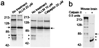
Figure 2.
Calpain cleavage of human and mouse htt. (a) Calpain inhibitors ALLN, ALLM, and calpeptin block formation of the proteolytic products of the human N-httcasp3 fragment. X57 cells were transfected and treated with inhibitors as described in Materials and Methods. Western blots (10 μg protein per lane) were probed with 2166 antibody. Arrows identify the level of the N-httcasp3 fragment, and arrowheads mark the level of the “C-terminal” portions of the calpain-cleaved fragments that are detected by antibody 2166. (b) Mouse brain homogenates treated with calpain II show increase in the levels of two N-htt fragments at arrowheads migrating at about 65 kDa and 55 kDa. The arrow identifies the level of the caspase-cleaved fragments in mouse htt. Blot was probed with Ab1. Twenty micrograms of protein was loaded per lane. The numbers beside blots in a and b are molecular mass markers.

