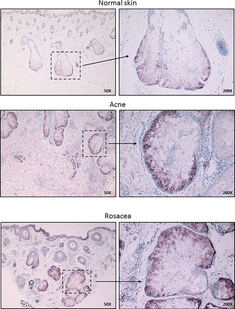
Fig 5. Immunohistochemical detection of Serum Amyloid A 1/2 marks activated sebocytes and reveals a structurally defined immune-competence in sebaceous glands.
Immunohistochemical detection of SAA1/2 in human sebaceous glands in control skin as well as in papulopustular acne and papulopustular rosacea. FFPE tissue samples were stained with rabbit monoclonal SAA1/2 antibody as described in the Materials and Methods. Note that besides the increased staining intensities observed in papulopustular acne and papulopustular rosacea samples, SAA1/2 positivity had a characteristic distribution within the sebaceous glands, localizing exclusively to the basal cell layers. Images are representative of at least 5 samples from each disease and each staining. Staining intensities were independent of age and gender. Sections were counterstained with methylene green. Original magnification: 50X, 200X.

