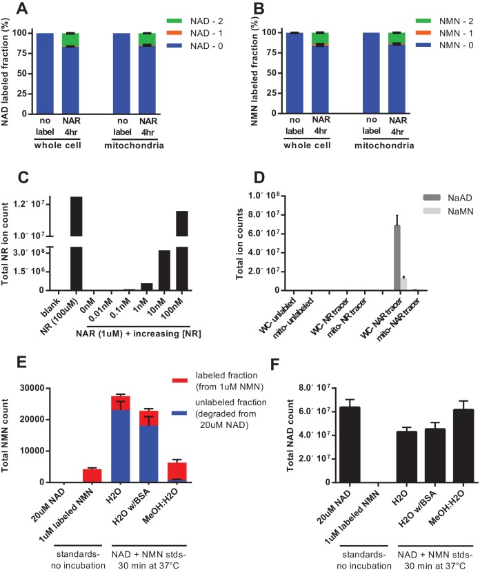
Figure 5. Nicotinic acid riboside is incorporated intact into mitochondrial NAD.
(A) Fractional labeling of NAD in C2C12 whole cell lysates and isolated mitochondria following 4 hr of incubation with doubly-labeled NAR. (N = 5). (B) Fractional labeling of NMN in C2C12 whole cell lysates and isolated mitochondria following 4 hr of incubation with doubly-labeled NAR. (N = 5). (C) Confirmation of the lack of NR contamination in NAR. 1 µM NAR was combined with increasing concentrations of NR (0–100 nM) to demonstrate that NR is absent in the NAR and readily detected by this methodology. (Single measurements). (D) Total ion counts for NAAD and NAMN in whole cell lysates and mitochondrial isolates from differentiated C2C12 cells treated with isotopically-labeled NR or NAR tracers for 4 hr. Results expressed as means ± SEM. (N = 3). (E) Incubation of NAD at 37°C in water, but not 80% methanol results in substantial degradation to NMN. Blue bars show unlabeled NMN resulting from degradation from a 20 µM NAD standard spike; Red bars indicate labeled NMN from spiked-in standard (1 µM, dual labeled). (N = 3). (F) NAD total ion count measured in parallel from same samples in (E). (N = 3).


