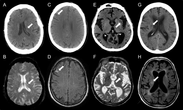
Figure 1.
Examples of misclassification of old infarcts between CT and MRI. A small lacunar infarct was just visible on CT (A) but below the size threshold for recording. The same lesion appeared larger on T2w MRI (B). A small cortical lesion missed on CT (C) was obvious on FLAIR MRI due to tissue signal changes (D). Low attenuation in the external capsule was recorded as an old infarct on CT (E) but was found to represent a cluster of EPVS on T2w MRI (F). A focal area of low attenuation observed on CT was identified as a noncavitated lacunar lesion (G); this was considered to be a WMH on FLAIR MRI (H). All assessments of old infarct agreement was based on data from 1 rater.

