
Figure 4. GluCer Converts Physiological α-Syn Conformers into Pathogenic Species.
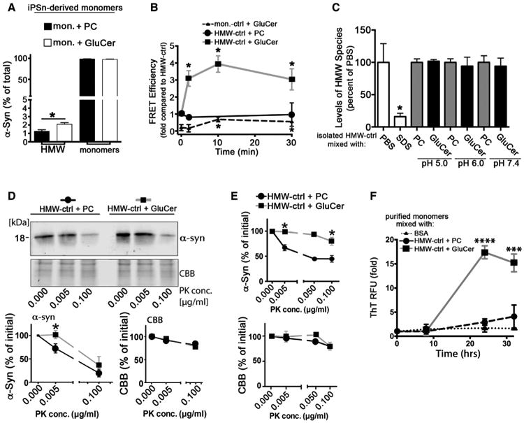
(A) iPSn-derived monomers isolated from control neurons were incubated with phosphatidylcholine (PC) as a control, and GluCer under quiescent conditions at pH 5.5, followed by SEC/ELISA to monitor conversion into HMW species (n = 4).
(B) Physiological conformers (HMW-ctrl or mon.-ctrl) were isolated from iPSns and mixed with PC or GluCer in vitro as in (A), followed by immuno-FRET analysis (n = 4, *p < 0.05 compared to time = 0).
(C) HMW-ctrl was isolated and mixed with PC or GluCer in vitro at pH 5.0, 6.0, or 7.4 for 30 min at 37°C followed by SEC/ELISA. SDS (0.1%) was used as a control to disrupt HMW conformers (n = 4).
(D) HMW-ctrl fractions from iPSns were treated with PC or GluCer as in (B) for 30 min, followed by PK digest/western blot (C-20)(n = 3). CBB was used as a loading control.
(E) HMW-ctrl from H4 cells was mixed with PC or GluCer in vitro and analyzed as in (D) (n = 3).
(F) HMW-ctrl from iPSns was incubated with BSA, PC, or GluCer in vitro as in (D), then mixed with purified recombinant monomers. Amyloid formation was monitored over time under quiescent conditions at pH 7.4 by ThT (n = 4). For all quantifications, values are the mean ± SEM (*p < 0.05, ***p < 0.0002,****p < 0.0001).