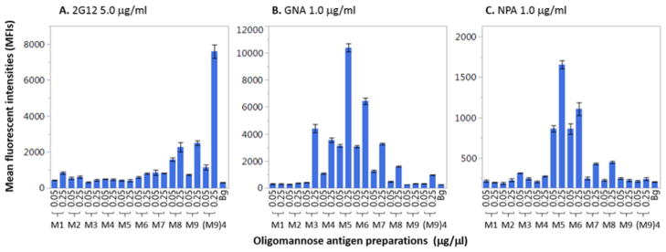
Figure 3.
Binding profiles of mannose-reactive proteins 2G12, GNA, and NPA to oligomannose–BSA conjugates. The glycoconjugates were spotted at 0.05 μg/μL and 0.25 μg/μL. Glycan-binding activities of antibody/lectin were shown as means of fluorescent intensities (MFIs) of triplicate microspots. Each error bar was constructed using one standard deviation from the mean of triplicate detections. The background (Bg) signal served as the negative control. The symbols M1 to M9 represent the neoglycoproteins ManGlcNAc2Asn-BSA to Man9GlcNAc2-BSA (Compounds 23–31).

