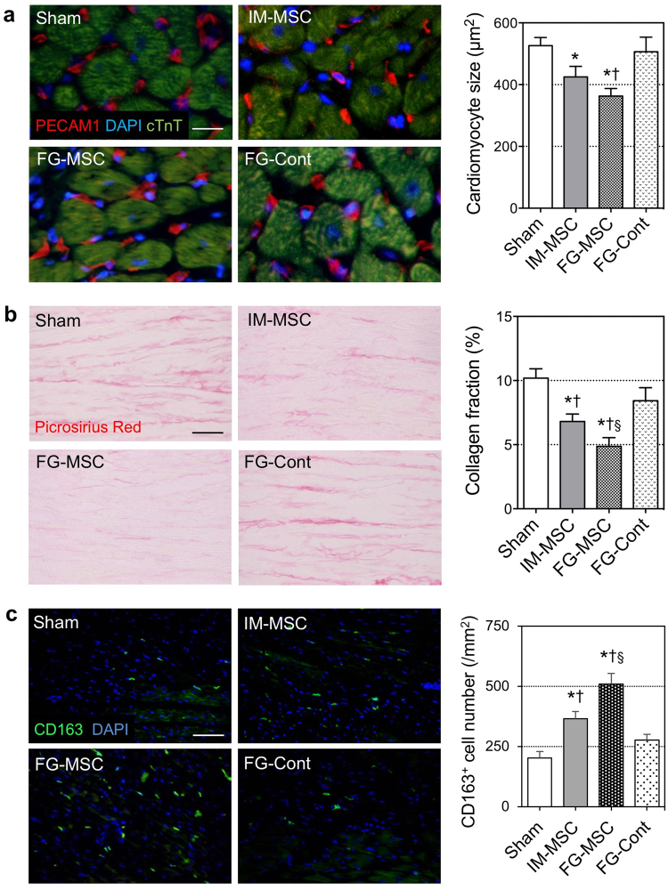
Figure 5.
Attenuated adverse ventricular remodeling after FG-aided, epicardial placement of MSCs in the ICM rat heart. (a) Cross-sectional area of cTnT cardiomyocyte was measured using immunohistolabeling samples at day 28 post-treatment. Scale bar = 50 μm. (b) Post-MI interstitial fibrosis in the peri-infarct area was assessed by picrosirius red staining at day 28 post-treatment. Scale bar = 100 μm. (c) Accumulation of CD163+ cells (green) in the myocardium at day 7 was most evident in the FG-MSC group. Scale bar = 100 μm. n = 6 hearts in each group. *p < 0.05 vs. the Sham group, † p < 0.05 vs. IM-MSC group. § p < 0.05 vs. FG-Cont group.

