Fig. 8.

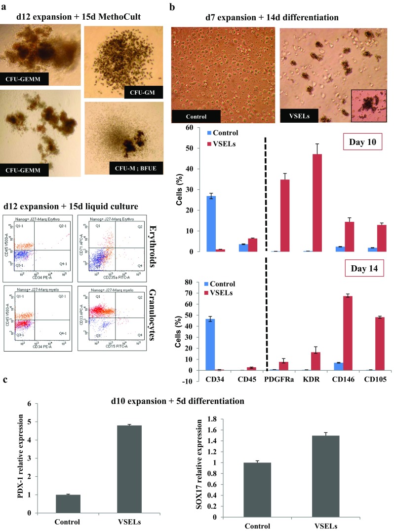
Expanded VSELs differentiation. a Hematopoietic colonies formation during 15 days in MethoCult from the sorted Lin-CD34 + CD45-NANOG+ VSELs which were previously expanded 12 days in culture medium containing UM171 (Upper panels). FACS analysis of hematopoietic cells formation from 15 days culture of Lin-CD34 + CD45-NANOG+ VSELs after 12 days expansion in culture medium containing UM171 (Lower panels). b The VSELs and control cells morphologies after 10 days of expansion and 14 days of mesoderm differentiation (upper panel). Phenotypic analysis by flow cytometry of PDGFRα, KDR, CD146 and CD105 mesoderm markers in VSELs and controls cells induced to differentiate in MV06™ media for 10 and 14 days. Bars represent average ± SD, n = 3 (*P < 0.05). c SOX17 and PDX-1 endodermic markers mRNA expression by real time RT-PCR of Lin-CD34 + CD45-CD133+ VSELs and control (CD133-) cells that were previously expanded for 10 days and induced to differentiate 5 days in endodermic medium.
