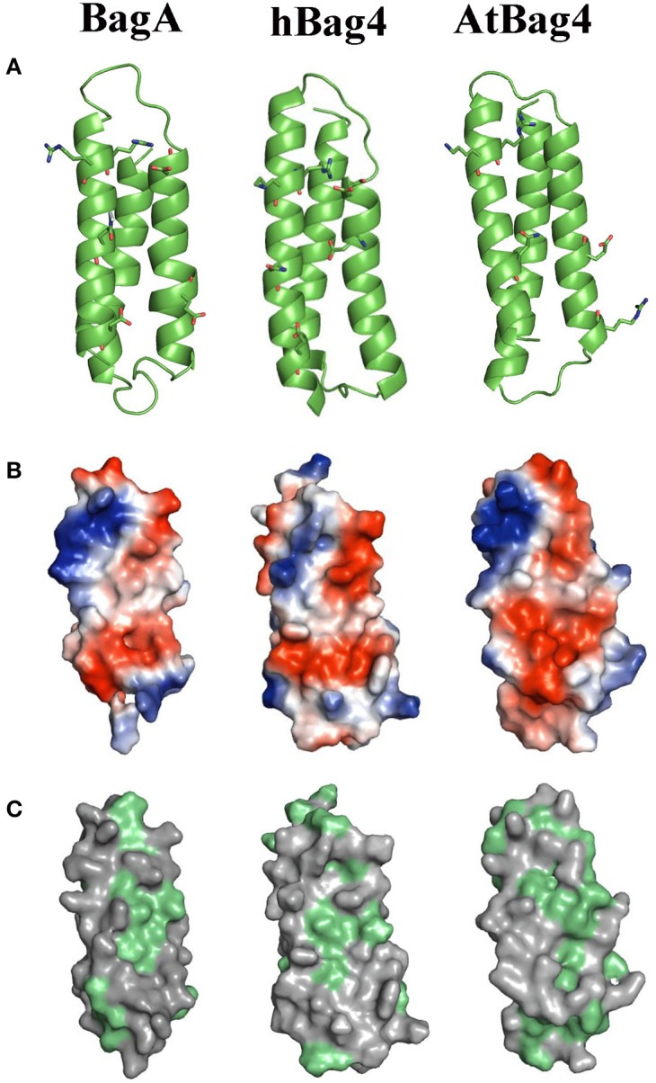
Figure 2.

Homology modeling, electrostatic potential, and hydrophobicity of BagA. (A) Ribbon representation of BagA BD, homolog model is shown along with solution NMR structures of AtBag4 (Protein data bank code: 4HWH) and hBag4 (Protein data bank code: 1M7K). Green ribbons represent three anti-parallel alpha helices with second and third helices facing forward. (B) The hydrophilic surface is colored based on the electrostatic potential (Negative –Red to Positive- Blue). The second and third helices are orientated toward the front similar to the ribbon representation. (C) The surface map of hydrophobic residues is shown; hydrophobic residues are highlighted in green.
