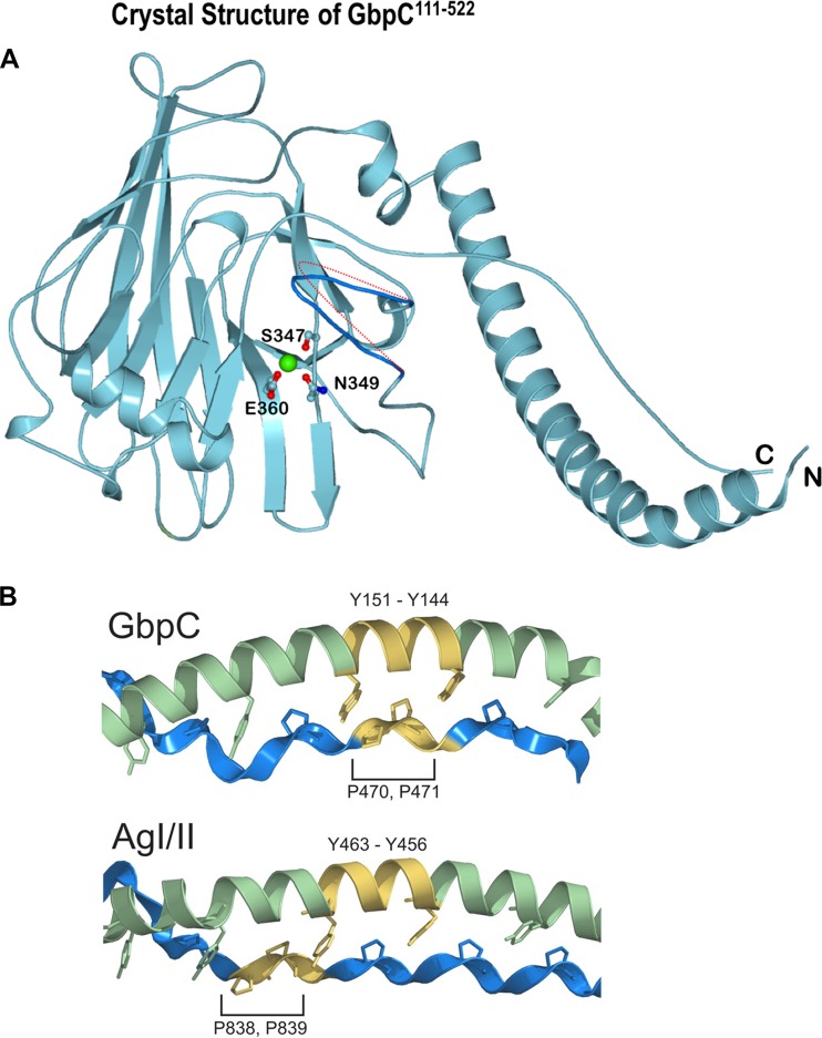
FIG 2.
(A) Crystal structure of GbpC111–522 with the lectin-like fold; the calcium binding site is highlighted. Three residues, Ser347, Glu360, and Asn349, form bipyramidal geometrical coordination for the calcium binding site. The loop region of residues 410 to 418 is highlighted in dark blue, and the potential movement of this loop region based on the B factors (see Table S2 in the supplemental material) is denoted by the dotted red line. (B) A closer examination of the A-P interaction on GbpC and AgI/II shows that there is a register shift in the interactions of the AxYxAx(L/V) motif on the A region and the PxxP motif on the P region that we had described earlier (3).

