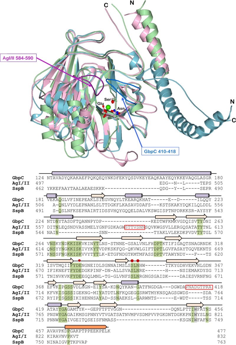
FIG 3.
The figure shows the structural superposition of the V regions of AgI/II (light pink), SspB (light green), and GbpC (light blue) and their structure-based sequence alignment with alpha-helices (purple cylinders), strands (beige arrows), and the PPII helix (saffron). The residues of the conserved calcium-binding site are highlighted (red dots on the sequence). The loop regions that hover over the calcium-binding site which can be imagined to emanate from two sides of a palm are highlighted in dark pink for AgI/II (residues 584 to 590) and dark blue for GbpC (residues 410 to 418; shaded red boxes in sequence). SspB does not display either of these two loop regions.

