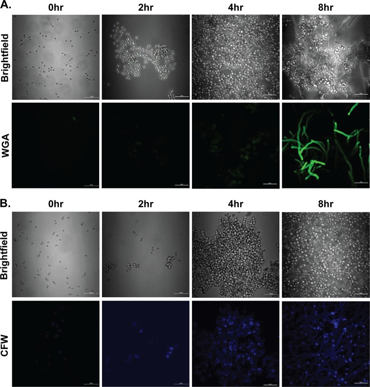
FIG 1.
Chitin is detected in increasing amounts as A. fumigatus conidia transition to hyphae. A. fumigatus conidia were resuspended in RPMI medium supplemented with 1% penicillin-streptomycin-glutamine and 10% heat-inactivated FBS and cultured for 0, 2, 4, and 8 h on glass slides at 37°C in a 5% CO2 incubator. For the 0-h time point, conidia were incubated for 1 h at room temperature to allow adherence. Adhered conidia were washed with PBS for 5 min and stained with Oregon green 488-conjugated wheat germ agglutinin at 37°C (A) or with calcofluor white at room temperature (B). Stained conidia were washed twice with PBS before mounting. 2D fluorescent images of stained conidia were imaged by using a Nikon A1 high-speed confocal laser scanning microscope with constant parameters across samples. Corresponding bright-field images are included to aid in the visualization of the culture. Representative images are shown.

