FIG 2.
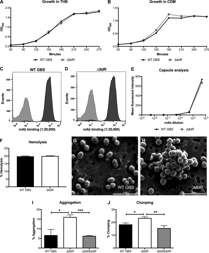
(A and B) Growth curves for WT GBS and the ΔltdR mutant in THB (A) and CDM (B) at 37°C. (C to E) Flow cytometry using serial dilutions of a monoclonal antibody (MAb) to the serotype III capsule to determine the presence of capsule in WT GBS (C) and the ΔltdR mutant (D). A monoclonal antibody to the serotype Ia capsule was used as the isotype control. (F) Hemolysis assay comparing the hemolysis of sheep blood cells by WT GBS and the ΔltdR mutant. Representative data from 1 of at least 2 independent experiments are shown. (G and H) Scanning electron microscopy images of WT GBS (G) and ΔltdR mutant (H) strains. (I) Aggregation assay comparing aggregation of WT GBS, the ΔltdR mutant, and the complemented strain in THB. (J) Clumping assay comparing clumping of the WT GBS, the ΔltdR mutant, and the complemented strain in THB containing 0.1% fibrinogen. *, P < 0.05; **, P < 0.005; ***, P < 0.0005.

