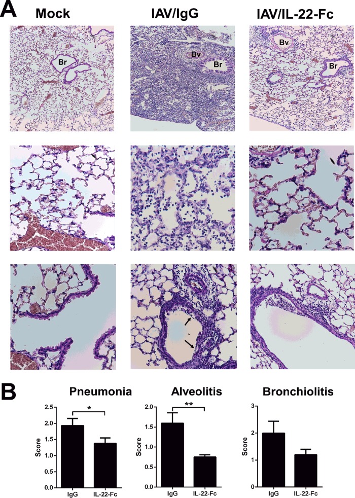
FIG 4.
Histological analysis of lung sections from IAV-infected (at 7 days postinfection) mice untreated or treated with IL-22. (A) Representative lung sections stained with hematoxylin and eosin are shown. Alveolar lesions (middle row) and perivascular and peribronchic infiltrates combined with lesions of alveolitis (bottom row) are shown. Arrows indicate denuded epithelia. Bv, blood vessel, Br, bronchiole. (B) Sections were scored blindly for levels of immunopathology. Data represent the means ± standard deviations (n = 5 mice/group). *, P < 0.05; **, P < 0.01 (Mann-Whitney t test).

