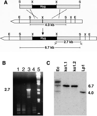
Figure 3.
Knockout of lpsA in Neotyphodium sp. Lp1. (A) Strategy for disrupting the internal 4.0-kb SalI fragment and for detecting the integration by PCR (symbols > and < indicate where primers LPKO-F and LPKO-R, respectively, anneal to prime amplification of a 2.7-kb fragment) and Southern blot hybridization (increased length of the disrupted SalI fragment is indicated). S = SalI; X = XhoI; E = EcoRI; Hyg = hygromycin resistance. (B) PCR product from primers LPKO-F and LPKO-R, indicating homologous recombination (lane 3 only) from spores of one of several transformants (lanes 1–4). Lane 5 = BstEII-digested bacteriophage λ DNA. (C) Southern blot probed with the 4.0-kb SalI fragment of N. lolii lpsA (used to direct the integration). Fragment sizes (in kb) are derived from BstEII digested bacteriophage λ DNA. Strain names are described in Results.

