. 2016 Apr 5;7(7):4016–4023. doi: 10.1039/c5sc03796d

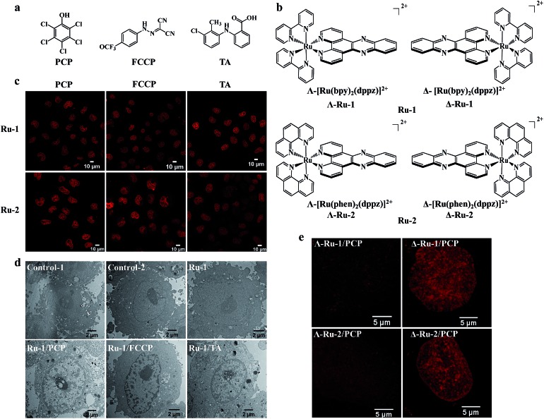
Fig. 2. Nuclear uptake of Ru(ii) complexes was enhanced by three structurally unrelated biochemical agents and the enantioselective imaging of live-cell nuclear DNA by the two chiral forms of Ru(ii)–dppz complexes. Chemical structures of the three biochemical agents (a) and the enantiomers of Ru(ii)–dppz complexes (b). (c) Confocal images of cells treated with Ru-1 (100 μM) or Ru-2 (100 μM) in the presence of PCP (300 μM) for 3 h in complete medium, Ru-1 (200 μM) or Ru-2 (500 μM) in the presence of FCCP (50 μM) for 1 h in serum-free medium, and Ru-1 (300 μM, 3 h) or Ru-2 (100 μM, 4 h) in the presence of TA (300 μM) in complete medium. (d) Cellular localization of Ru-1, characterized by transmission electron microscopy (TEM) in the absence or presence of PCP, FCCP, and TA for 3 h. Negative control cells stained with nothing (control-1) or solely with osmium tetroxide (control-2); cells incubated with Ru-1 (1 mM) alone, Ru-1 (300 μM)/PCP (300 μM), Ru-1 (500 μM)/FCCP (100 μM), Ru-1 (500 μM)/TA (300 μM) and additionally stained with osmium tetroxide. (e) Dramatic differences were observed between the two chiral forms (Δ and Λ) of Ru-1 and Ru-2 in live-cell nucleus DNA imaging, with Δ-enantiomers showing much brighter emission intensity inside the nucleus compared to Λ-enantiomers. Structured illumination microscopy images of cells incubated with Δ-Ru (100 μM) or Λ-Ru (100 μM) in the presence of PCP (300 μM) in cells for 3 h.

Fig. 2