. 2016 Apr 5;7(7):4016–4023. doi: 10.1039/c5sc03796d

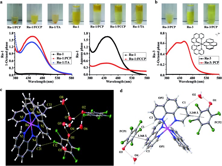
Fig. 3. Evidence for the formation of neutral and lipophilic ion-pair complexes between Ru(ii) complexes and the three biochemical agents. (a) Precipitation of Ru-1 and PCP, FCCP, TA, in aqueous buffer solution; partition studies of Ru-1 (100 μM) between 1-octanol and aqueous phases (tris–HCl buffer, 10 mM, pH 7.4) in the absence or presence of PCP (1 mM), FCCP (1 mM), and TA (1 mM). (Note: because both FCCP and Ru-1 have similar strong absorption spectra, to avoid their interference with each other, the spectra were obtained in aqueous phase in the case of FCCP). (b) Precipitation between Ru-3 and PCP in aqueous buffer solution, and partition studies of Ru-3 (100 μM) in the absence or presence of PCP (1 mM). (c) Crystal structure of the ion pair complex [Ru(phen)3]2+(PCP)2 by ORTEP drawing with 30% thermal ellipsoids, the H-bonds are indicated by dotted lines. (d) Crystal packing diagram showing stacking interactions between [Ru(phen)3]2+ cation and PCP anion within the [Ru(phen)3]2+(PCP)2 complex. The black dotted lines indicate the π···π interactions between the PCP ring and the parallel phen ring; the red dashed line indicates the cation–anion C–H···(H2O)···O hydrogen bonds; the green dashed line indicates the cation–anion C–H···Cl hydrogen bond. The other solvent molecules were omitted for clarity.

Fig. 3