. 2016 May 4;7(7):4004–4008. doi: 10.1039/c6sc01463a

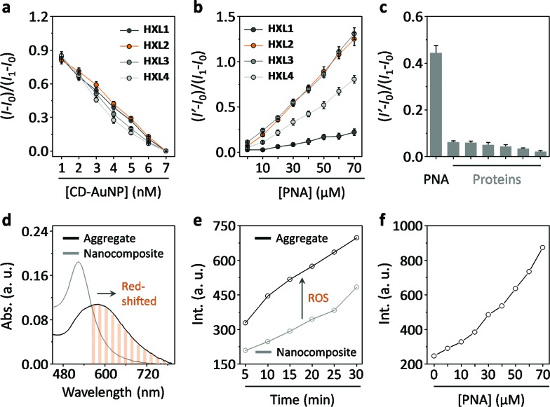
Fig. 2. (a) Concentration-dependent fluorescence quenching of HXLs (0.56 μM) in the presence of increasing CD-AuNP. (b) Concentration-dependent fluorescence recovery of HXLs@CD-AuNP (0.56 μM/7 nM) in the presence of increasing peanut agglutinin (PNA). (c) Fluorescence enhancement of HXL2@CD-AuNP (0.56 μM/7 nM) in the presence of different proteins (30 μM, from left to right: pepsin, wheat germ agglutinin, ribonuclease, bovine serum albumin, concanavalin A and Lens culinaris lectin) (I1, I0, I and I′ are the fluorescence intensity of HXL, HXL@CD-AuNP, HXL with CD-AuNP of a certain concentration and HXL@CD-AuNP with a protein of a certain concentration, respectively). (d) UV-vis absorbance of HXL2@CD-AuNP (0.56 μM/7 nM) in the absence and presence of PNA (70 μM). (e) Reactive oxygen species (ROS) production after irradiation (600 nm) of HXL2@CD-AuNP (0.56 μM/7 nM, composite) in the absence and presence of PNA (30 μM, aggregate) with time. (f) Reactive oxygen species (ROS) production of HXL2@CD-AuNP (0.56 μM/7 nM) with increasing PNA after irradiation (600 nm) for 10 min.

Fig. 2