Figure 1.

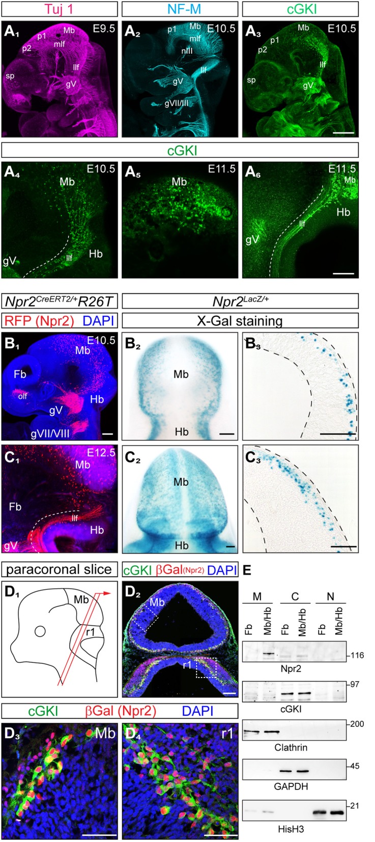
Co-expression of cGKI and Npr2 in the dorsal midbrain and rhombomere 1 at early embryonic stages. (A) Analysis of the pattern of expression of cGKI in cells and axon tracts of the mesencephalon at E9.5, E10.5, and E11.5. For comparison, axons tracts labeled by Tuj1 (A1) or neurofilament (A2) from confocal z-stacks are shown. (A3-A6) Anti-cGKI staining shows the localization of cGKI positive cell somata in the midbrain and descending axons. Scale bar in (A1-A3), 200 μm; in (A4-A6), 50 μm. (B) Detection of Npr2-expressing cells using the Npr2-CreERT2-line in combination with the R26T reporter line (R26-LSL-tdTomato) [in red, counterstained with an antibody to red fluorescent protein (RFP)] or using Npr2-LacZ mice by X-Gal staining. Lateral view of a head of a whole mount (B1; z-stacks), dorsal view (B2) and a sagittal section (B3) of the midbrain are shown at E10.5. Scale bar for (B1–B3), 100 μm. (C) Expression of Npr2 monitored using the reporter line R26T (activated by Npr2-CreERT2) or by using the reporter Npr2-LacZ. Lateral view of a head in a whole mount preparation (C1; z-stacks), dorsal view (C2) and sagittal section of the midbrain are shown at E12.5. Scale bar for (C1–C3), 100 μm. (D) Co-localization of Npr2 and cGKI in a neural population in the dorsal midbrain and rhombomere 1 of cryostat sections at E12.5. Localization of Npr2 was demonstrated by anti-β-galactosidase staining of the Npr2-LacZ reporter mouse line. (D3) (mesencephalon) and (D4) (rhombomere 1) show higher magnifications of squares indicated in (D2). The scheme illustrates the orientation of sectioning (D1). gV, trigeminal ganglion; gVII, geniculate ganglion; gVIII, ganglion of the vestibulocochlear nerve; Hb, hindbrain; llf, lateral longitudinal fascicle; Mb, midbrain; mlf, medial longitudinal fascicle (rostral to the prosomere 1-mesencephalic boundary); nIII, oculomotor nerve; ofl, olfactory placode; p1, prosomere 1; p2, prosomere 2; r1, rhombomere 1; sp, secondary prosencephalon. Scale bar in (D2–D4) 100 μm. (E) Western blotting of subcellular fractions of the forebrain and midbrain/hindbrain extracts (E11.5) using antibodies to Npr2 or cGKI. Loading control was obtained by antibodies to clathrin heavy chain (crude membrane fraction), GAPDH (cytoplasmic fraction) or histone H3 (nuclear fraction). M, crude membrane fraction; C, cytoplasmic fraction; N, nuclear fraction. Molecular mass markers are indicated at the right of the panels in kD.
