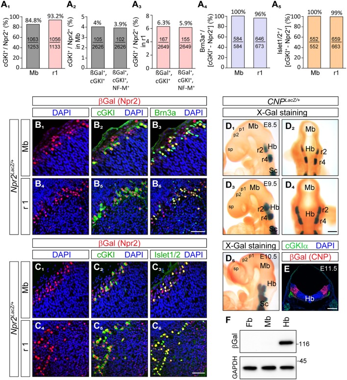
Figure 2.
Brn3a and Islet1/2 co-localize with Npr2/cGKI in midbrain and rhombomere 1 and the ligand CNP is localized in the hindbrain but absent from the midbrain and forebrain. (A1) Percentage of cGKI-positive cells of Npr2-expressing cells in the mesencephalon and in rhombomere 1 at E12.5. (A2,3) Percentage of Npr2(β-Gal)/cGKI-positive or Npr2(β-Gal)/cGKI/neurofilament-positive cells of total midbrain or rhombomere 1 cells. Total cells were identified by DAPI. Anti-β-galactosidase staining was performed using the Npr2-LacZ reporter mouse line to monitor Npr2-expressing cells. (A4,B) Co-localization of Brn3a with Npr2(β-Gal)/cGKI-positive cells in the midbrain or rhombomere 1 at E12.5. (A5,C) Co-localization of Islet1 with Npr2(β-Gal)/cGKI-positive cells in the midbrain or rhombomere 1 at E12.5. The Npr2-LacZ reporter line was used to monitor expression of Npr2 in (B, C) by using an antibody to β-galactosidase. Scale bar for (B1-C6), 50 μm. (D) Localization of CNP at E8.5, lateral (D1) and dorsal (D2) view of whole mounts. Expression analysis was done with the CNP-LacZ reporter mouse line using X-Gal staining. Localization of CNP at E9.5, lateral (D3) and dorsal (D4) view. (D5) Localization of CNP at E10.5 lateral view. Scale bar for (D1–D5) 30 μm. (E) Cross section of the hindbrain at the level of rhombomere 2 at E11.5 indicating that CNP is localized throughout all layers of the hindbrain. Anti-β-galactosidase staining of the CNP-lacZ reporter mouse line was used to monitor CNP. Scale bar, 250 μm. Hb, hindbrain; Mb, midbrain; Pros, prosencephalon; r2, rhombomere 2; r4, rhombomere 4; Sc, spinal cord. (F) Western blot of extracts of hindbrain, midbrain, and forebrain from CNP-LacZ reporter mice at E13.5 using anti-β-galactosidase to monitor expression of CNP. Equal loading was demonstrated by anti-GAPDH. Molecular mass markers are indicated at the right of the panel in kD.

