Figure 3.
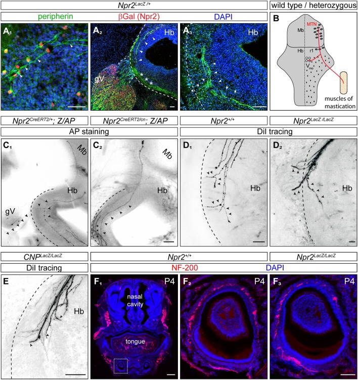
In the absence of Npr2 or CNP MTN axons fail to form Y-shaped bifurcations in rhombomere 2 of the hindbrain. (A1) Peripherin co-localizes with Npr2-positive cells in the midbrain and (A2,3) is found in descending MTN axons which extend in lateral regions of the hindbrain (arrow heads). Trigeminal axons are also positive for peripherin which grow at the lateral border of the hindbrain (asterisks). Anti-β-galactosidase staining monitors Npr2 expression in Npr2-LacZ reporter mouse. Scale bar 50 μm. (B) Scheme of bifurcating MTN afferents. Hb, hindbrain; Mb, midbrain; MTN, mesencephalic trigeminal neuron; r1, rhombomere 1; Vmo, trigeminal motor nucleus. (C1) An Npr2-positive axon originating in rhombomere 1 which bifurcates in rhombomere 2 from a heterozygous Npr2 mutant. One arm is leaving the hindbrain via the root of the trigeminus demarcated by arrowheads growing toward V3 of the trigeminus (mandibular region). The other branch stays within the hindbrain to grow to Vmo. Another afferent originating in the midbrain had not reached the bifurcation zone (transgenic sparse labeling by alkaline phosphatase staining using the Z/AP reporter mouse line method). (C2) A MTN axon (arrow heads) bypassing the bifurcation zone without generating a peripheral branch from an Npr2 knockout mutant (sparse labeling by using Z/AP). Trigeminal axons which are entering the hindbrain and which also do not bifurcate in global Npr2 knockouts are marked by stars. Scale bar for (C1, 2) 200 μm. (D1) DiI tracing of MTN axons in the wild type hindbrain at the level of rhombomere 2. Two bifurcating axons are indicated by arrowheads. In 5 control embryos (Npr2+/+, CNP+/+ and Npr2CreERT2/+;Z/AP) 15 bifurcating MTN afferents were observed. No non-bifurcating afferents were found (see also Table 1). (D2) In the absence of Npr2 MTN afferents do not bifurcate and either exit or extend further in the hindbrain. Single non-bifurcating axons and fascicles of axons are shown. Scale bars in (D1,2) 50 μm. (E) In the absence of the ligand CNP no bifurcating MTN axons were observed at the level of the dorsal root of the trigeminus. (D2,E) In 4 homozygous mutants (CNPLacz/LacZ, Npr2LacZ/LacZ, and Npr2CreERT2/cn;Z/AP) 40 non-bifurcating and 4 bifurcating MTN afferents were observed (see also Table 1). Scale bar, 50 μm. (F) Neurofilament staining of a transversal section of a tooth at P4 show less axons in the absence of Npr2. (F1) Overview of a transversal section of the mouth at P4; neurofilament staining of wild type (F2) and Npr2 knockout (F3). Scale bar in (F1), 500 μm; in (F2, 3), 100 μm. The square in (F1) denotes the region enlarged in (F2).

