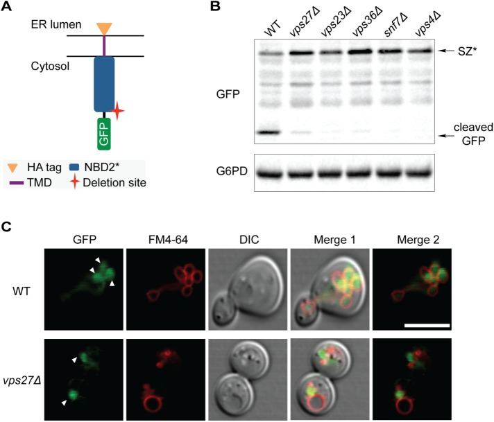
FIGURE 4:
SZ* is sorted into the vacuole through the MVB pathway. (A) Predicted topology of GFP-tagged SZ*. The cytosolic residence of GFP allows for analysis of MVB delivery to the vacuole. (B) GFP cleavage from GFP-SZ* in the wild-type and indicated mutant strains was determined by immonoblotting. G6PD serves as a loading control. (C) The cellular localization of SZ* in both wild-type and vps27Δ cells was determined by fluorescence microscopy. FM4-64 marks the vacuolar membrane. Arrowheads denote the lobes of the vacuole (Bohovych et al., 2016) and the prevacuolar compartment (bottom). Scale bar: 5 μm.

