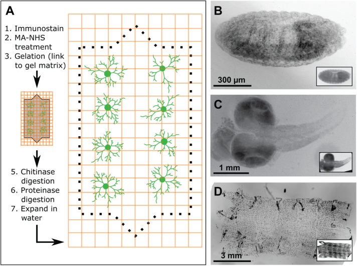
FIGURE 1:
Isotropic expansion of Drosophila tissues for fluorescence microscopy. (A) ExM workflow. (B–D) Correlative pre- and postexpansion imaging of Drosophila tissue. Drosophila embryos (B), larval brains (C), and larval body walls (D) were stained with 4′,6-diamidino-2-phenylindole (DAPI) and imaged both before and after expansion. Preexpansion (inset) and postexpansion images are shown at the same magnification, such that postexpansion images are approximately four times larger than the corresponding preexpansion images.

