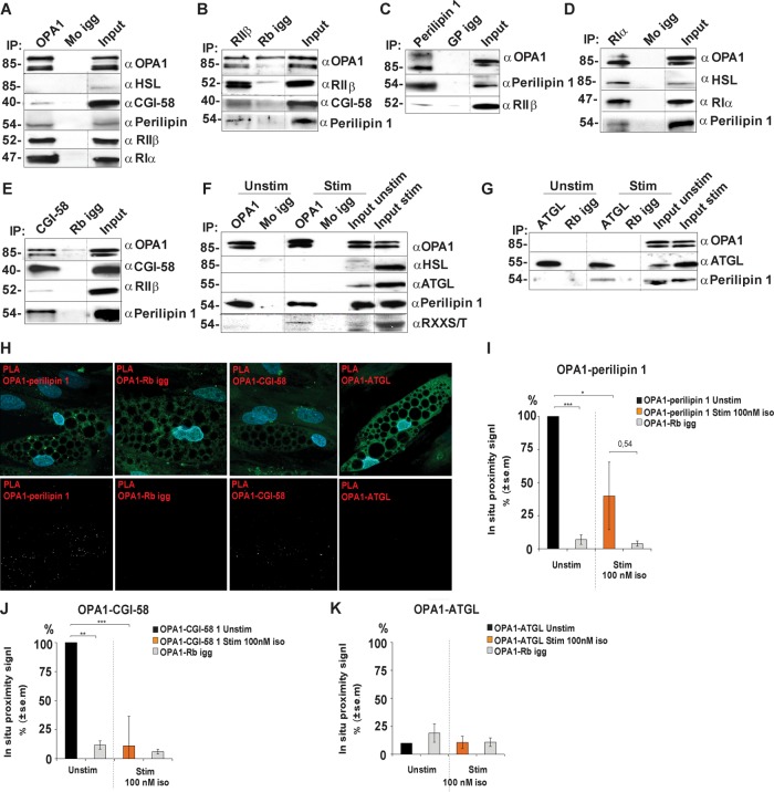
FIGURE 3:
Association of OPA1 with perilipin and PKA in adipocyte-differentiated hASCs. Lysates from hASC-derived adipocytes were immunoprecipitated with antibodies to OPA1 (A), RIIβ (B), perilipin 1 (C), RIα (D), and CGI-58 (E) and the appropriate igg control (Mo/Rb/GP igg). Precipitates were analyzed for the presence of the indicated protein interaction partners. Lysates from adipocytes incubated in the absence or presence of 10 nM isoproterenol were subjected to immunoprecipitation for OPA1 (F) and ATGL (G) and blotted for presence of the indicated protein interaction partners. Dotted lines indicate images merged from the same gel (different exposure time for input lysate) (n = 3 donors). In situ proximity studies using PLA for the OPA1-perilipin 1, OPA1-CGI-58, and OPA1-ATGL associations (red, top and bottom rows), lipid droplet visualization (green, top row), and DAPI (blue, top row) in hASC-derived adipocytes (H). Scale bars: 10 µm. Statistical analysis of in situ proximity experiments as in H of hASC incubated in the absence or presence of 100 nM of isoproterenol for 20 min and stained for OPA1-perilipin 1 (I), OPA1-CGI-58 proximity signal (J), and OPA1-ATGL proximity signal (K); *p < 0.05, **p < 0.005, ***p < 0.0005. Fifty cells were counted from each condition and for every donor (mean ± SEM, n = 3 donors).

