FIGURE 3:

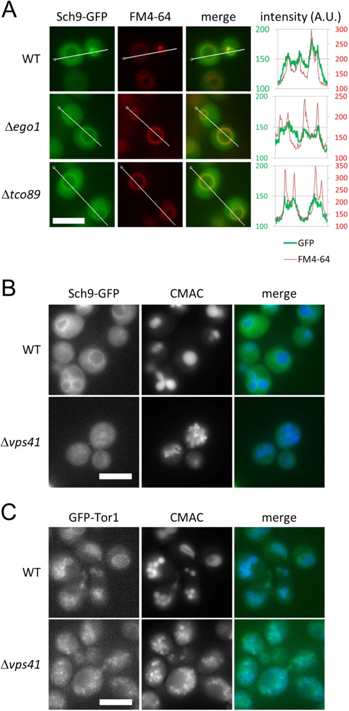
The HOPS complex is necessary for normal localization of Sch9. (A) Representative images of WT (yet120), ∆ego1 (yet691), and ∆tco89 (yet727) cells expressing Sch9-2GFP from the SCH9 promoter encoded on a centromeric plasmid. Cells at mid–log phase grown in SDC medium were stained with FM4-64 as a vacuolar marker. The signal intensities of Sch9-2GFP and FM4-64 along the indicated lines were measured by softWoRx software. Scale bar = 5 µm. (B) Representative images of WT (yet120) and ∆vps41 (yet732) cells expressing Sch9-2GFP. Cells at mid–log phase grown in SDC medium were stained with CMAC as a vacuolar marker. Scale bar = 5 µm. (C) Representative images of WT (SKY374-A) and ∆vps41 (yet665) cells expressing Tor1-GFP. Cells at mid–log phase grown in SDC medium were stained with CMAC as a vacuolar marker. Scale bar = 5 µm.
