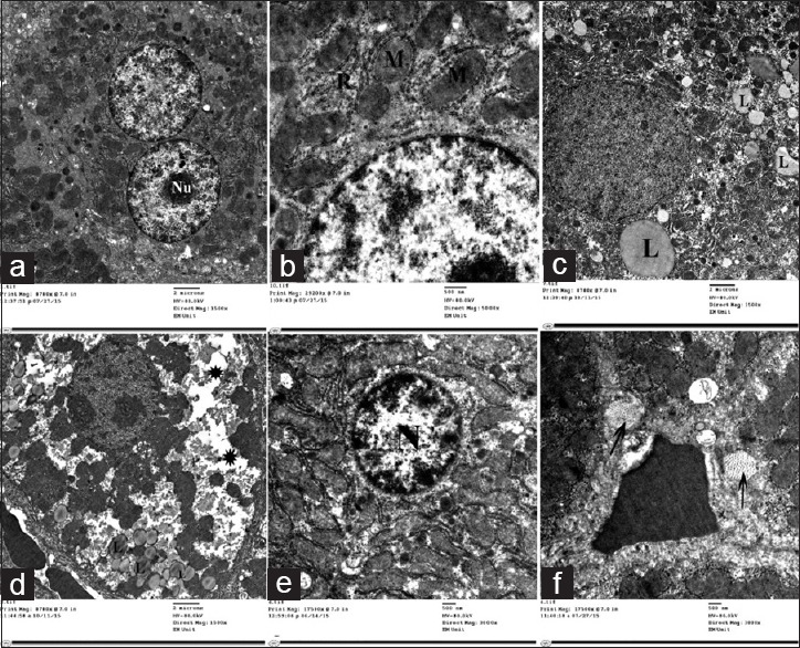
Figure 11.

Electron micrographs of liver ultrathin section of Group IV showing (a) Multiple hepatocytes some of them are binucleated with prominent nucleolus (Nu), (b) Preserved ultrastructural picture including mitochondria (M) and rough endoplasmic reticulum (R). (c) Others showing many lipid droplets (L). (d) Rarefied cytoplasm (*). (e) Nuclei with condensed chromatin (N) and (f) Bundles of collagen fibers (arrow) in between the hepatocytes
