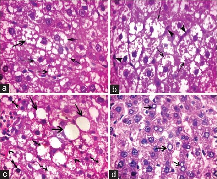
Figure 5.

Photomicrograph of liver sections of Group III showing hepatocytes with variable cytoplasmic vacuolation. (a) Multiple small vacuoles (arrow) (b) large coalesced vacuoles (arrow) and multiple small cells with ovoid nuclei arranged in rows in between the hepatocytes (arrowheads). (c). Ballooned hepatocytes containing one large vacuole (arrow), some hepatocytes with darkly stained nuclei (biffed arrow), fragmented nuclei (curved arrow), and lysed nuclei (waved arrow). (d) Some hepatocytes with vacuolated nuclei (arrow) (H and E, ×1000)
