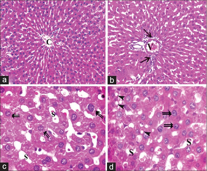
Figure 6.

Photomicrographs of liver sections of Group IV showing apparently mild dilatation and congestion of central vein (C), portal vein (V) blood sinusoids (S) with mild cellular infiltrations (arrow) around components of a portal tract. Most of hepatocytes appear normal (double arrow) and few cells containing some cytoplasmic vacuoles (arrowhead) (H and E, [a and b] × 400 [c and d] × 1000)
