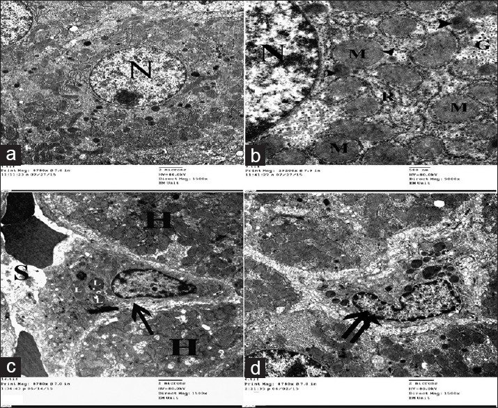
Figure 9.

Electron micrographs of liver ultrathin sections of control group showing (a) Polyhedral-shaped hepatocyte-containing large rounded nucleus (N) with extended chromatin and prominent nucleolus. (b) The cytoplasm-containing abundant mitochondria (M), rER (R), glycogen granules (G), and multiple peroxisomes (arrowhead). (c) Space of Disse between hepatocytes (H) and blood sinusoids (S) containing HSCs cell (arrow) with multiple lipid with droplets (L). (d) Kupffer cell (double arrow) appears as irregular shaped cell with indented nucleus and multiple cytoplasmic dense bodies
