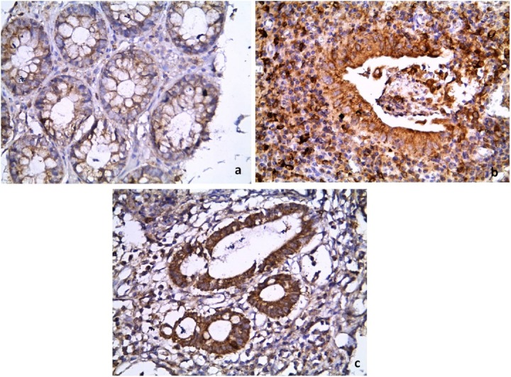
Figure 1.
Comparison of the immunohistochemical pattern of COX-2 between: (a) the control, that shows weak cytoplasmic epithelial staining; and (b, c) the UC group, that shows strong cytoplasmic staining in epithelial cells and inflammatory cells. (Immunoperoxidase: 400×).
COX-2 = cyclooxygenase-2; UC = ulcerative colitis.

