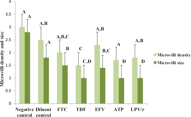
Fig. 3.

Changes observed in microvilli density and size in HCS-2 cells following treatment with antiretroviral drugs. There was a significant difference observed between the different groups (p<0.05). Drug treatment reduced the size and density of microvilli although the changes were unique to each drug treatment. TDF, ATP and LPV/r treatment showed the most reduction in microvilli size and density in comparison to controls and other drug treatments. Bar graphs not connected by the same letter (per characteristic) are significantly different, Tukey Kramer Post Hoc analysis.
