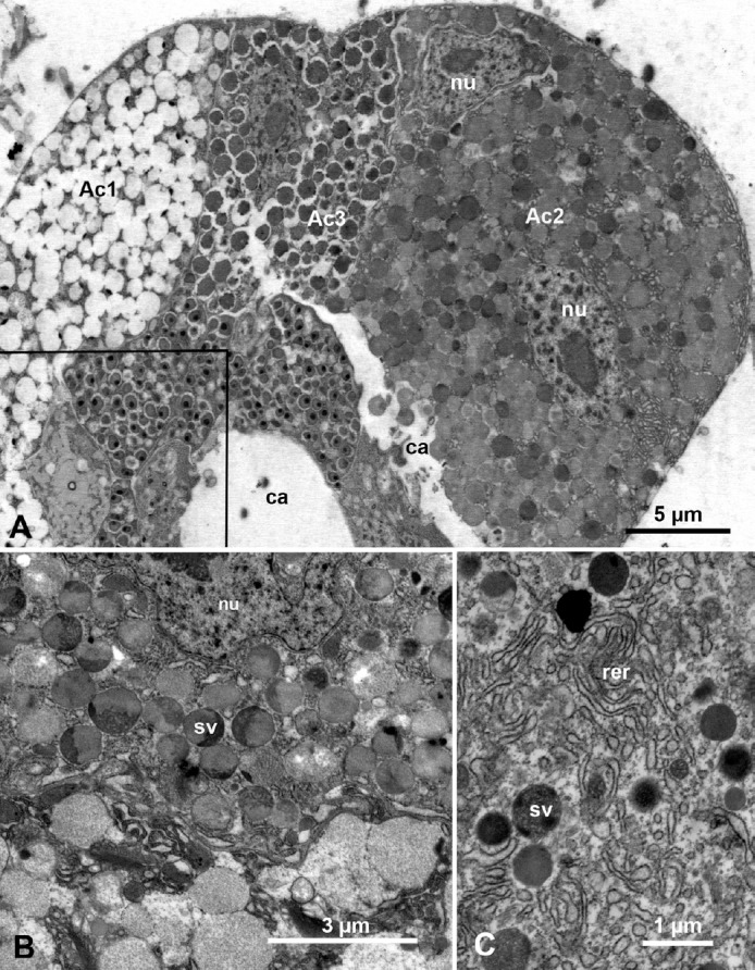
Fig. 2.

Ultrastructure of the principal salivary gland in Diaphorina citri (A) nymphs and (B, C) adults. (A) Part of the principal salivary gland showing three types of acini: Ac1 with electrolucent secretory vesicles, Ac2, with both semiopaque and electron-dense secretory vesicles, and Ac3 with mainly electrondense secretory vesicles; boxed area is shown at higher magnification in Figure 4A; note the intercellular canaliculi (ca) between acini and the two nuclei (nu) in acinus Ac2. (B) Part of acinus Ac2 showing the nu and secretory vesicles (sv) with various electron-dense or semiopaque material. (C) Electron-dense sv and surrounding cytoplasm, rich with rough endoplasmic reticulum (rer).
