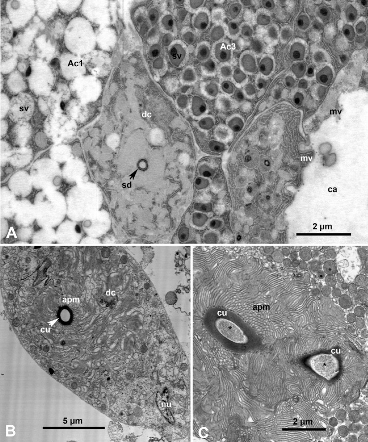
Fig. 4.

Ultrastructure of the salivary glands and salivary ducts in Diaphorina citri. (A) Higher magnification of the boxed area in Figure 2A, showing two types of acini (Ac1 and Ac3), salivary duct cells (dc), salivary duct (sd), and a large intercellular canaliculum (ca) lined with microvilli (mv). (B, C) Cross-sections in the salivary ducts, the lumen of which is lined with a cuticle (cu), surrounded by elaborate infoldings of the apical plasma membrane (apm) of dc; asterisks indicate semiopaque material (putative salivary secretions) in the duct lumen; nu, nucleus.
