
Fig. 6.
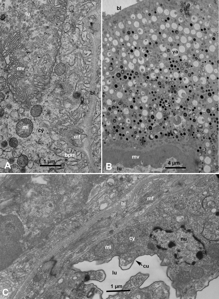
Ultrastructure of the midgut, Malpighian tubule and hindgut in Diaphorina citri adults. (A) Higher magnification of part of an epithelial cell of the midgut, showing basal lamia (bl), muscle fibers (mf), invaginated basal plasma membrane (bpm), cytoplasm (cy), mitochondria (mi), microvilli (mv), and lumen (lu). (B) Part of an epithelial cell of a Malpighian tubule, lined with fine mv, with various-sized cytoplasmic vacuoles (va) either apparently empty, partially full or full of dark excretory material; bl, basal lamina. lu, lumen. (C) Part of the epithelium of the hind gut showing circular mf, bl, nucleus (nu), mi, cy, and cuticular lining (cu) around the lu.