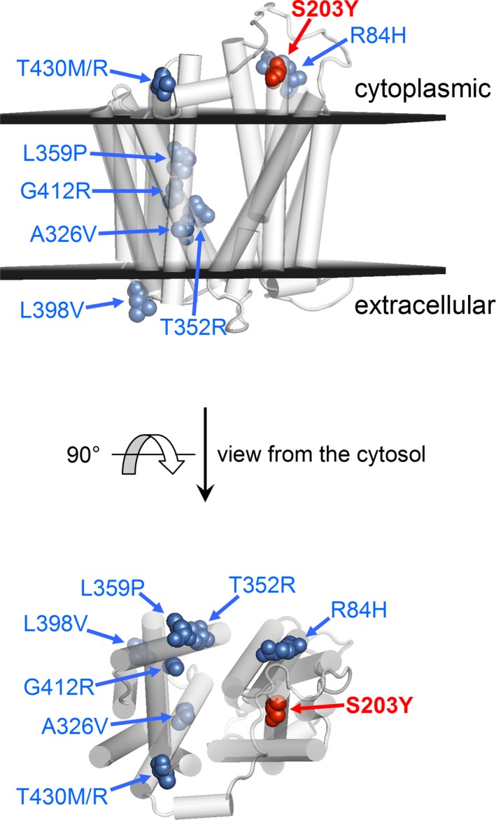
Figure 3.

Homology models of FLVCR2 (residues 86–491) based on GlpT di E. coli, PDB 1PW4 model (first on the left in Figure 2c). Known mutants are shown as colored spheres and marked by blue arrows. Substrates are transported across the membrane through the central part of the protein along the direction perpendicular to the figure plane of the cytosol view
