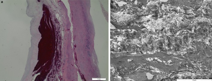
Figure 2.

Histology and electron microscopy of the aorta of patient III‐1. Left: hematoxylin‐stained section of the aorta showing dissection and hemorrhage as well as myxoid media degeneration. On the left: ultrastructure of the aorta with smooth muscle cell degeneration, but without characteristic morphologic features of Ehlers‐Danlos‐syndrome, Marfan‐syndrome, or a heritable connective tissue disorder with known ultrastructural aberrations
